 |  |
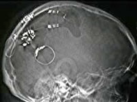
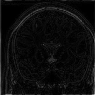
The second image is the result of computerized axial tomography (better known in the medical world by the acronym CAT) on the same subject. In this case, the parallel X-ray beam is located on a uniform network segment. This segment is rotated about an axis on the subject slowly until complete 360 \u200b\u200bdegrees, and each pause, take the intensities received after the broadcast. As a result, we get a picture square, each of its columns providing information on emissions at different angles. In the picture above is collected 180 breaks, and the parallel beam consisted of 100 bulbs.
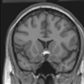
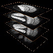
But this image is not easy to interpret, is where one has to use the power of mathematics, more specifically techniques An & # 225; advanced mathematical analysis and numerical analysis addressing the reversal of the Radon transform. After a series of numerical calculations (based on algorithms that after more a century are still being perfected), the doctor will get the following picture:
 |
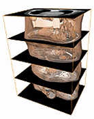
But overall investment Radon transform is a very costly process, mainly by the volume of information accumulated-note that this operation must be repeated as many times as necessary down and up the front axle in order to obtain an accurate three-dimensional image inside the patient. Not all medical institutions can afford the maintenance adquisicióny CT equipment to obtain the best possible definition, and the scanners used in most health centers offer some approximation errors & # 243; n derived from these technological deficiencies.
 |  |
 |
Technorati Tags: IMA , math, math , tomography, Radon transform
0 comments:
Post a Comment